
Tumor staining is a crucial technique used in neurosurgery to enhance the precision of tumor identification and resection. It helps surgeons clearly differentiate between healthy and diseased tissue, which is particularly important in brain and spinal surgeries, where surrounding neural structures are highly delicate. By providing a more detailed visualization of tumor boundaries, tumor staining optimizes the safety and effectiveness of the surgical procedure.
The Mechanism of Tumor Staining in Neurosurgery
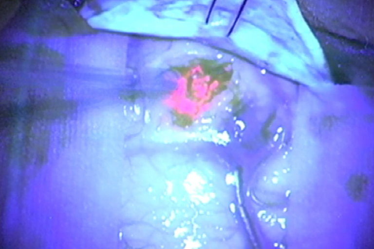
Tumor staining involves the application of special dyes or contrast agents to a tumor or tissue during surgery, which enables it to be visualized more clearly. These dyes or agents may be absorbed preferentially by tumor cells, causing the tumor to appear brightly colored under specific lighting or with the aid of fluorescence microscopes. Some stains are used to differentiate tumor cells from normal tissue, while others highlight specific tumor characteristics such as blood vessels or areas of increased metabolic activity.
The most common method of tumor staining in neurosurgery includes:
Fluorescent Staining: This technique involves the use of fluorescent dyes, such as 5-aminolevulinic acid (5-ALA), which are absorbed by tumor cells and cause them to fluoresce under certain light conditions. 5-ALA is particularly useful in glioma surgeries, as it accumulates in tumor cells and allows surgeons to distinguish malignant tissue from surrounding healthy brain matter.
The Role of Tumor Staining in Neurosurgery
The primary function of tumor staining is to provide a clearer, more detailed visual representation of the tumor and its surrounding tissue. In the intricate environment of neurosurgery, where tumors are often in close proximity to vital brain regions or spinal cord structures, precise removal is essential to minimize the risk of damage. Tumor staining helps achieve this by delineating tumor boundaries, revealing hidden tumor remnants, and assisting in the identification of areas at risk for potential neurological damage.
Tumor staining serves several specific purposes in neurosurgery:
Improved Tumor Visualization: Tumors, especially those in the brain, can appear similar to surrounding tissue, making it difficult to distinguish between normal and abnormal areas. Tumor staining amplifies the tumor’s visibility, allowing for more precise tumor removal.
Identification of Malignant Tissue: Certain stains specifically bind to tumor cells, making them stand out clearly against healthy tissue. This is particularly beneficial in surgeries involving malignant tumors such as gliomas, where removing as much cancerous tissue as possible while sparing normal neural structures is crucial.
Advantages of Tumor Staining in Neurosurgery
Enhanced Surgical Precision: Tumor staining significantly improves the surgeon’s ability to accurately visualize the tumor and its borders, which is crucial for maximizing tumor removal and minimizing the risk of neurological damage. In the case of gliomas, which infiltrate surrounding brain tissue, the ability to differentiate tumor from healthy tissue is critical for achieving a complete resection while preserving brain function.
Lower Risk of Residual Tumor: One of the major challenges in neurosurgery is ensuring that the entire tumor is removed. Tumors can have irregular borders and extend into surrounding tissues, making it difficult to fully excise them. Tumor staining helps ensure that no tumor tissue is left behind, reducing the likelihood of recurrence.
Faster and More Efficient Surgery: By providing real-time, highly detailed visuals of the tumor, tumor staining can reduce the time spent identifying tumor boundaries during surgery. This leads to quicker and more efficient procedures, which can be especially important in high-risk surgeries where prolonged operation time increases the risk of complications.
Improved Prognosis and Outcomes: By enabling surgeons to achieve a more complete resection, tumor staining can improve the chances of long-term success and reduce the likelihood of recurrence. This contributes to better overall patient outcomes, including improved survival rates and enhanced quality of life following surgery.
Better Visualization of Invasive Tumors: Some tumors, like glioblastomas, invade surrounding healthy tissue, making them difficult to completely remove without damaging critical brain structures. Tumor staining provides a clearer delineation of the invasive regions, allowing for more thorough removal and reducing the risk of leaving behind malignant cells.