
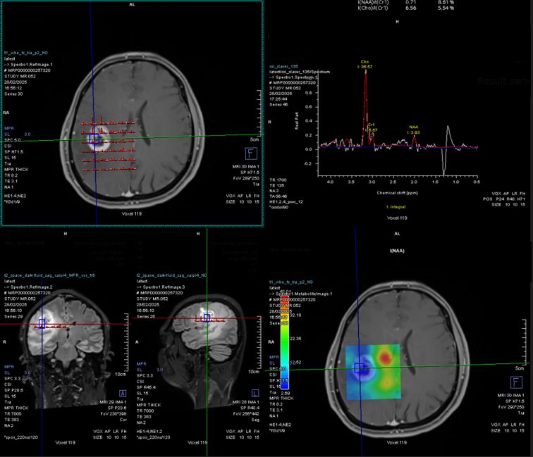
MR Spectroscopy (Magnetic Resonance Spectroscopy) is an advanced imaging technique that measures the chemical composition of tissues, providing critical information about the biochemical environment of the brain. Unlike standard MRI, which primarily focuses on anatomical structures, MRS analyzes the metabolic profile of brain tissue by detecting the concentrations of various metabolites, such as choline, creatine, N-acetylaspartate (NAA), and lactate. These metabolites can indicate changes in cellular metabolism that are associated with various neurological conditions, including tumors, infections, and degenerative diseases. MR Spectroscopy is a clinical application of nuclear magnetic resonance spectroscopy, a technique originally used in chemistry to analyze molecular structures.
Applications
1.Differentiating Between Brain Tumors:
- Tumor Characterization: One of the primary uses of MRS in neurosurgery is in the differentiation of brain tumors. MRS can provide crucial information about the metabolic activity of a lesion, helping to distinguish between malignant and benign tumors. Malignant tumors typically show elevated levels of choline (a marker of cellular turnover), decreased levels of N-acetylaspartate (NAA) (a marker of healthy neuronal tissue), and the presence of lactate or lipids (which indicate tissue necrosis or hypoxia).
- Tumor Type and Grade: Magnetic resonance spectroscopy brain, helps to refine the understanding of the tumor type (e.g., gliomas, meningiomas, metastases) and its grade (e.g., low-grade vs high-grade glioma) based on the unique biochemical signatures of the tumor tissue. This metabolic information can guide the surgical team in determining the extent of resection and planning the surgical approach.
MR Spectroscopy © ENI
2.Assessing Tumor Response to Treatment:
- Monitoring Treatment Efficacy: MRS is valuable for monitoring how brain tumors respond to treatments such as chemotherapy, radiotherapy, or targeted therapy. By comparing pre- and post-treatment metabolic profiles, it helps assess whether the tumor is shrinking, stable, or showing signs of recurrence. For example, a decrease in choline levels and an increase in NAA levels can indicate a positive treatment response, whereas elevated lactate levels may signal a lack of response or tumor progression.
- Post-Surgical Monitoring: After tumor resection, MRS can be used to evaluate the residual tumor tissue and surrounding brain areas for any signs of tumor recurrence or metabolic abnormalities that might not be visible on conventional MRI or CT scans. This is especially important in high-grade gliomas, where there may be small areas of infiltrating tumor that are not easily identified through anatomical imaging alone.
3.Differentiating Between Tumors and Other Lesions:
- Lesion Differentiation: MRS is also useful in distinguishing between tumors and other types of brain lesions, such as abscesses, infections, vascular malformations, or neurodegenerative diseases. For example, brain abscesses typically show elevated lactate levels and a reduced presence of NAA, which helps differentiate them from gliomas or metastatic tumors that have different metabolic signatures.
- Non-Tumor Lesions: In cases of stroke, infection, or neurodegeneration, MRS can help clarify the nature of the lesion by assessing metabolic changes in the affected tissue. It can help identify areas of ischemia (low NAA) or infection (high lactate), which can influence the surgical or medical management of the condition.
4.Preoperative Planning for Tumor Resection:
- Guiding Surgical Approach: MRS provides important metabolic data that can influence the surgical strategy for tumor resection. For instance, if a tumor is located near critical functional areas of the brain (such as the motor cortex or language areas), MRS can help assess the tumor’s proximity to these regions and assist in surgical planning to minimize damage to healthy tissue. It can also highlight areas where tumor cells may have infiltrated normal brain tissue, which helps determine the extent of safe resection.
- Aiding in Navigational Systems: The metabolic data from MRS can be integrated into neuronavigation systems (such as stereotactic navigation) during surgery, helping the surgeon navigate toward areas of abnormal metabolism, ensuring that the resection is as complete as possible while preserving critical brain function.
5.Postoperative Evaluation and Follow-Up:
- Monitoring for Recurrence: After brain surgery, particularly tumor resection, MRS is useful for detecting early signs of tumor recurrence. This is critical in cases of aggressive tumors like glioblastoma, where recurrent tumor growth often occurs at the margins of the resection site. MRS can detect metabolic changes in the tissue that suggest tumor growth before it becomes visible on routine MRI scans.
- Evaluation of Brain Tissue Health: MRS can also help evaluate the health of surrounding brain tissue after surgery, providing insight into whether the surrounding neural tissue is recovering or showing signs of damage, such as necrosis or edema.
6.Evaluation of Metabolic Changes in Neurodegenerative Diseases:
- In addition to its use in tumor surgery, MRS can help evaluate metabolic changes in neurodegenerative diseases like Alzheimer’s, Parkinson’s, or multiple sclerosis. While these conditions may not require surgical intervention, MRS can help differentiate between neurodegenerative changes and other lesions in the brain, and can also provide useful data for assessing the progression of these diseases.
Advantages in Neurosurgery
Metabolic Profiling of Brain Tumors:
- MRS provides a non-invasive method to assess the metabolic characteristics of brain tumors, enhancing the ability to distinguish between different types of tumors. This information aids in tumor characterization and helps guide surgical planning by identifying malignant versus benign lesions and assessing their potential infiltrative nature.
2.Improved Surgical Decision-Making:
- By providing a detailed metabolic profile of tumors and surrounding tissue, MRS can guide the surgeon in making critical decisions about the extent of resection. In particular, it helps to identify areas of active tumor tissue that may not be visible on standard imaging, allowing for a more complete resection while avoiding damage to healthy brain areas.
3.Monitoring Treatment Response:
- MRS is invaluable in assessing how well brain tumors are responding to chemotherapy, radiotherapy, or other treatments. This real-time feedback helps oncologists and neurosurgeons adapt treatment plans based on the metabolic activity of the tumor, ensuring that patients receive the most effective treatment.
4.Differentiating Between Tumors and Other Brain Lesions:
- MRS helps to differentiate tumors from other lesions, such as abscesses, vascular malformations, and infarctions, based on their unique biochemical signatures. This reduces the likelihood of misdiagnosis and ensures that the patient receives the appropriate treatment or surgical intervention.
5.Non-Invasive and Complementary Tool:
- MRS is a non-invasive procedure that can be performed alongside standard MRI, offering a complementary method for assessing brain tissue. It provides additional metabolic data that can aid in diagnosis and surgical planning without the need for invasive procedures like biopsy. This practical imaging technique is a clinical realization of nuclear magnetic resonance spectroscopy definition, tailored specifically for neurologic application.