
Neuro-Ultrasonography Techniques
Neuro-ultrasonography can be used in various ways during neurosurgery, depending on the specific clinical need. Some common applications of intraoperative ultrasonography include:
Brain Tumor Surgery: During tumor resections, intraoperative neuro-ultrasonography helps surgeons identify tumor margins and their relationship to critical neural structures, such as motor areas and sensory pathways. This ensures that tumor removal is as complete as possible while minimizing damage to surrounding healthy tissue.
pre-operative
post-operative
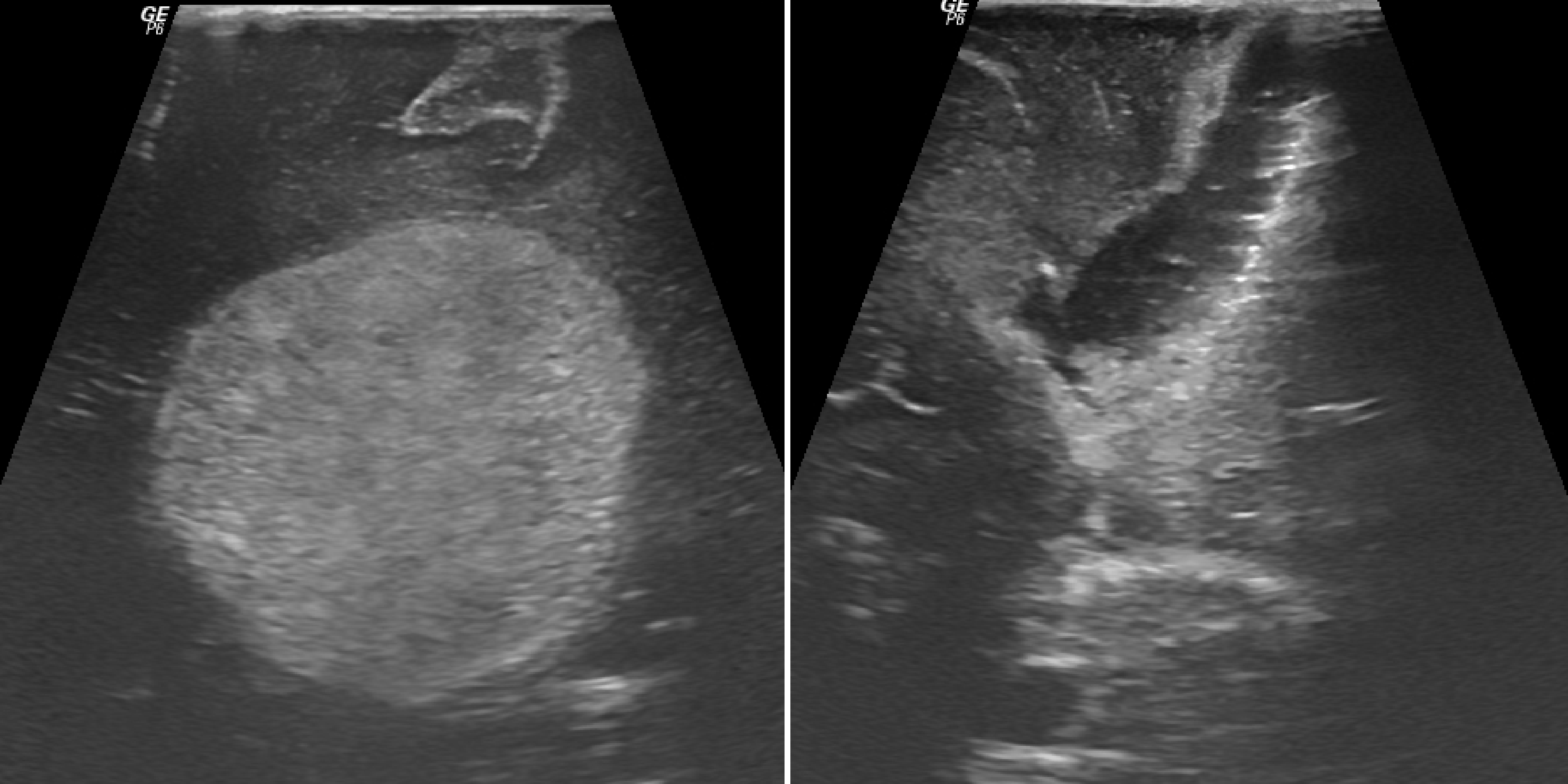
Neuroultrasonography © ENI
Spinal Surgery: In spinal cord surgeries, intraoperative neuro-ultrasonography provides real-time imaging of the spinal cord and nerve roots, enabling precise identification of the spinal structures. This helps reduce the risk of nerve damage and enhances surgical outcomes in spinal decompression or tumor removal.
Vascular Surgery: During neurosurgeries involving blood vessels, such as aneurysm repairs or arteriovenous malformation (AVM) resections, neuro-ultrasonography is used to visualize blood flow, assess vessel integrity, and guide surgical interventions.
Hydrocephalus and Ventricular Surgery: In procedures where the cerebrospinal fluid (CSF) dynamics are involved, such as shunt placement or ventriculostomies, neuro-ultrasonography helps assess the ventricular system in real time, ensuring proper placement and function of devices.
Advantages of Intraoperative Neuro-Ultrasonography in Neurosurgery
Intraoperative neuro-ultrasonography offers numerous advantages that contribute to the safety, precision, and success of neurosurgical procedures. These benefits include:
Real-Time Imaging: The ability to visualize neural structures and surrounding tissues in real time enables surgeons to make immediate, informed decisions during surgery. This helps reduce the likelihood of complications and enhances surgical accuracy.
Minimization of Neural Damage: By providing clear images of critical neural pathways, intraoperative neuro-ultrasonography assists in preserving motor, sensory, and other vital functions during brain and spinal surgeries. This reduces the risk of postoperative neurological deficits and enhances recovery.
Enhanced Surgical Precision: Neuro-ultrasonography offers high-resolution imaging, which allows the surgeon to precisely target areas of concern. Whether it’s tumor resection, spinal cord decompression, or vascular intervention, this technique improves the overall precision of the surgery.
Support for Complex Procedures: In complex surgeries, where the risk to neural pathways is high, neuro-ultrasonography provides a crucial support tool for decision-making. Surgeons can track the progress of the procedure, ensuring that all critical structures are preserved.
Real-Time Feedback for Multidisciplinary Teams: Neuro-ultrasonography provides visual data that can be used by the entire surgical team, including the neurosurgeon, anesthesiologist, and neuro-ultrasonography technician. This collaboration ensures that all team members are aligned in their approach to minimizing risk and improving outcomes.
Improved Postoperative Outcomes: The accuracy and effectiveness of intraoperative neuro-ultrasonography contribute to better postoperative recovery. By ensuring that critical neural structures are preserved during surgery, patients experience fewer complications and recover more quickly.