Neurosurgery requires extreme precision, as it involves operating on some of the most intricate and delicate structures within the human body. Surgeries on the brain, spinal cord, and nerves demands exceptional skill and attention to detail. Over time, innovations in technology have enabled surgeons to examine and intervene in deeper, smaller, and more complex structures. A key advancement in making micro-surgeries possible is the surgical microscope.
The Role of Microscopes in Neurosurgery
The advent of neurosurgical microscope in neurosurgery revolutionized the field, making it possible to visualize previously inaccessible structures. First introduced in the mid-20th century, microscopes allowed surgeons to perform micro neurosurgery, offering a detailed view of the smallest tissues without damaging sensitive nerve structures. The use of microscopes has not only made these surgeries safer but also shortened recovery periods.
With surgical microscopes, surgeons can clearly visualize both large areas and microscopic structures such as tiny blood vessels, nerves, and tumors. This is particularly critical when operating on the complex and delicate structures within the brain and spinal cord, where precise intervention is paramount.

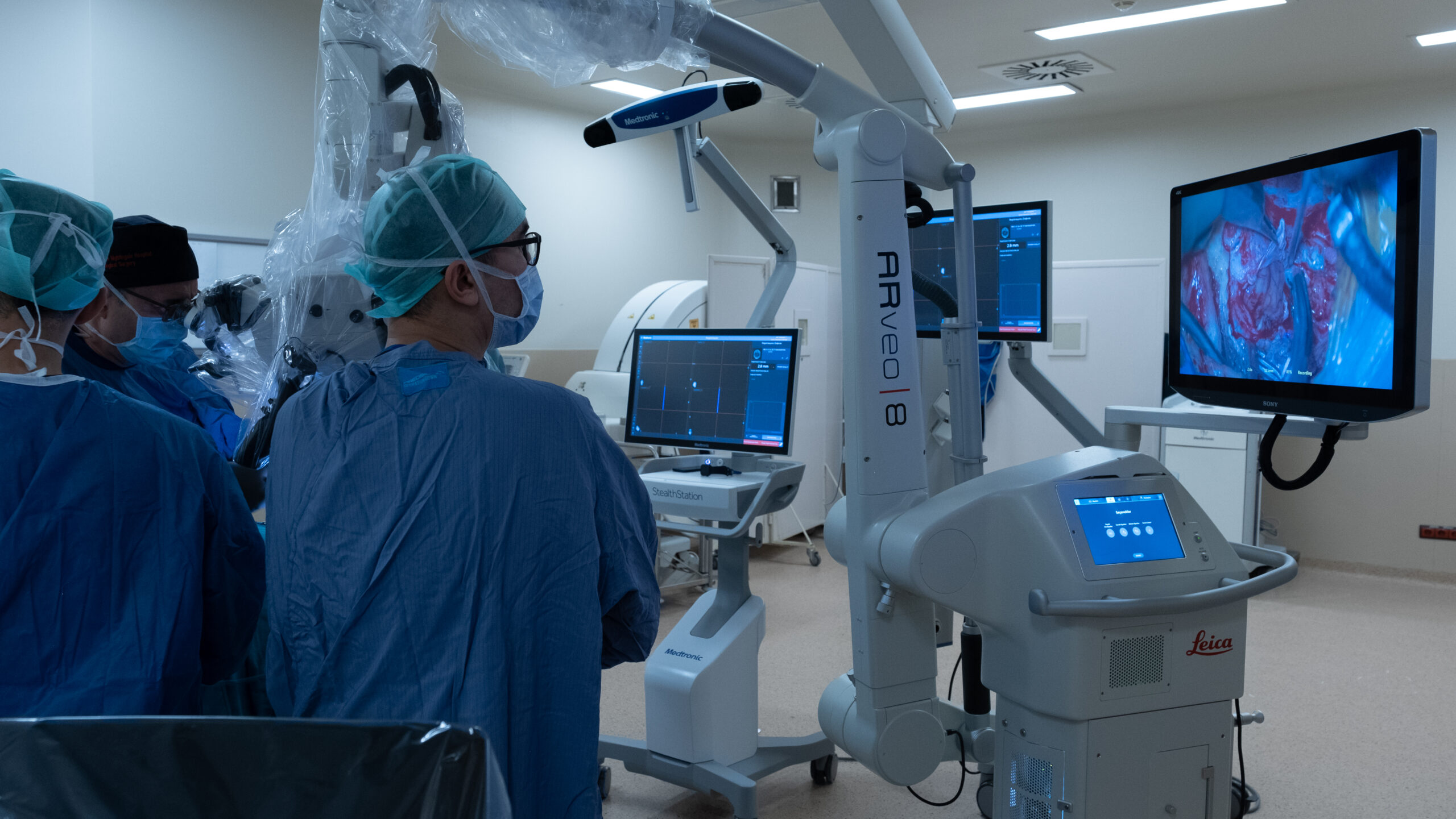
Surgical Microscope © ENI
M. Gazi Yaşargil and the Evolution of Microsurgery
The most influential figures in micro neurosurgery is Prof. M. Gazi Yaşargil, often hailed as the “father of modern neurosurgery.” Yaşargil’s groundbreaking work has had a lasting impact on the field, particularly his advancements in the application of surgical microscopes. His contributions have significantly enhanced the precision and safety of neurosurgical operations.
Yaşargil is credited with refining microsurgical techniques and pioneering the use of surgical microscopes in delicate procedures such as brain tumor removals, vascular malformations, and aneurysm repairs. His approach, combining meticulous techniques and the surgical microscope, enabled the preservation of vital neural structures that would otherwise have been difficult to locate and protect. This method has improved surgical outcomes while reducing risks of complications such as nerve damage and bleeding after surgery. His work laid the foundation for the advanced micro neurosurgery techniques used today.

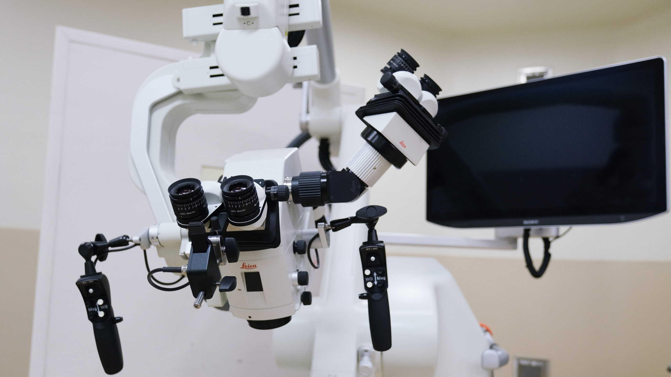
Surgical Microscope © ENI
How Micro Neurosurgery Works
Micro neurosurgery is performed using optical devices (surgical microscopes) and micro instruments. This surgical technique requires great precision when working on sensitive structures such as the brain and spinal cord. The use of a microscope allows surgeons to focus on smaller areas and clearly see critical structures such as blood vessels and nerves.
During micro neurosurgery, microscopes provide not only enhanced vision but also a wide field of view, allowing for more controlled and precise movements. The microscope offers the surgeon a magnified, detailed view of the surgical area, helping to preserve nerve structures and blood vessels while minimizing potential complications.
Advantages of Modern Surgical Microscope in Neurosurgery
- High-Quality Imaging and Precision: Microscopes, particularly high-resolution models, allow surgeons to clearly see fine structures. This makes it possible to perform micro-surgery with the utmost precision.
- Advanced 3D Imaging: Microscopes provide three-dimensional imaging, which enhances the surgeon’s depth perception and allows them to better identify and understand the anatomical structures.
- Safer Procedures: The use of microscopes allows surgeons to visualize small blood vessels and nerves without causing damage. This helps preserve nerve tissue and reduce complications.
- Ergonomic Use and Suitability for Long Procedures: The ergonomic design of microscopes allows surgeons to perform lengthy surgical procedures more comfortably. The adjustable design ensures surgeons can find the most efficient working position.
- Integrated Digital Systems: Modern microscopes are integrated with digital imaging and video systems, enabling surgeons to monitor the patient’s condition in real-time during the operation. This provides the opportunity to assess and optimize the procedure when needed.
